
A novel approach to understanding multiscale biogeochemical processes.
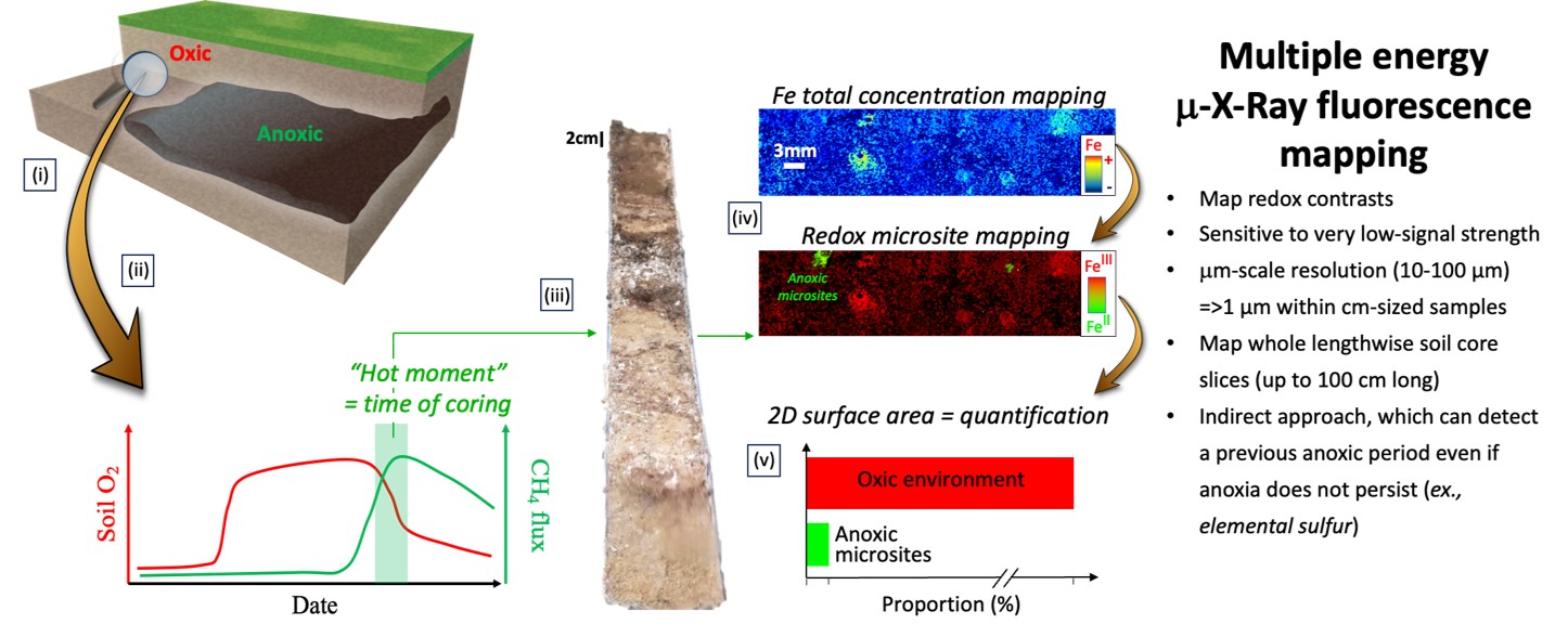
Image courtesy of Noël et al. (2024) Frontiers in Environmental Chemistry, 5:1329887.
Image Caption: Step-by-step method for detecting and quantifying hidden anoxic microsites from natural samples.
The Science
Soils play a crucial role in regulating Earth’s life-supporting functions, but biogeochemical process heterogeneities (=microsites) have yet to be incorporated into models of soil biogeochemistry. Small-scale redox microsites may significantly influence the cycling and release of nutrients and contaminants, which are important at larger scales. In this paper we developed a new approach to identify and quantify the spatially varying redox gradients arising from anoxic microsites (small zones deprived of oxygen) in generally oxygen-rich soils. These microsites contribute to nutrient and contaminant fluxes to and from soils but are typically not considered in bulk assessments due to their small size.
The Impact
This method paper provides a significant advancement in addressing the scientific need to identify and quantify redox microsites in soils and sediments. These investigations revealed direct evidence of anoxic microsites in predominantly oxic soils, such as those from the toeslope of a mountainous watershed, where anaerobic conditions would typically not be expected. Our approach offers the community a unique opportunity to detect and quantify anoxic microsites, thus providing the tools needed for future research to generate reliable experimental data that can be incorporated into numerical models predicting the biogeochemical cycling of nutrients and contaminants at the ecosystem scale.
Summary
Only a few studies have observed/characterized anoxic microsites in undisturbed soils, primarily, because soil is opaque and microsites require μm-cm scale resolution over cm-m scales. To address the challenge of detecting, quantifying and characterizing anoxic microsites in soils, we combine synchrotron X-ray fluorescence imaging (μ-XRF) and X-ray absorption near-edge structure (μ-XANES) spectroscopy. This approach enables 2-D mapping of the spatial distributions of total concentration, oxidation states, and chemical forms, of trace level elements that are indicative of current (e.g., FeIIS) or previous (e.g., presence of elemental sulfur, distinct depletion of solid-phase Fe and/or Mn, or accumulation of S and/or U) anaerobicity in natural soil core slices (up to 100 cm long). In this proof-of-concept study, we tested and developed a systematic data processing approach able to discriminate the presence of redox microsites at 1–100μm scale resolutions. Then, spatial distribution of FeII and FeIII species from full, natural soil core slices from a toeslope soil of a mountainous watershed (East River, CO) was mapped, revealing direct evidence of anoxic mm-sized microsites in a predominantly oxic soil.
Contact
Vincent Noël, SFA Science Lead
STAFF Scientist, SLAC National Accelerator Laboratory
Eoin L. Brodie, Watershed Function SFA LRM
Lawrence Berkeley National Laboratory
Funding
Funding was provided by the U.S. Department of Energy (DOE) office of Biological and Environmental Research (BER), Environmental System Sciences Division, through its support of the SLAC part of the Watershed Function Science Focus Area (SFA) under Contract No. DE- AC02-76SF00515.
Publications
Vincent Noël, Kristin Boye, Hannah Naughton, Emily Lacroix, Meret Aeppli, Naresh Kumar, Scott Fendorf, and Samuel Webb (2024) X-ray chemical imaging for assessing redox microsites within soils and sediments. Frontiers in Environmental Chemistry, 5:1329887.
[DOI: 10.3389/fenvc.2024.1329887]